

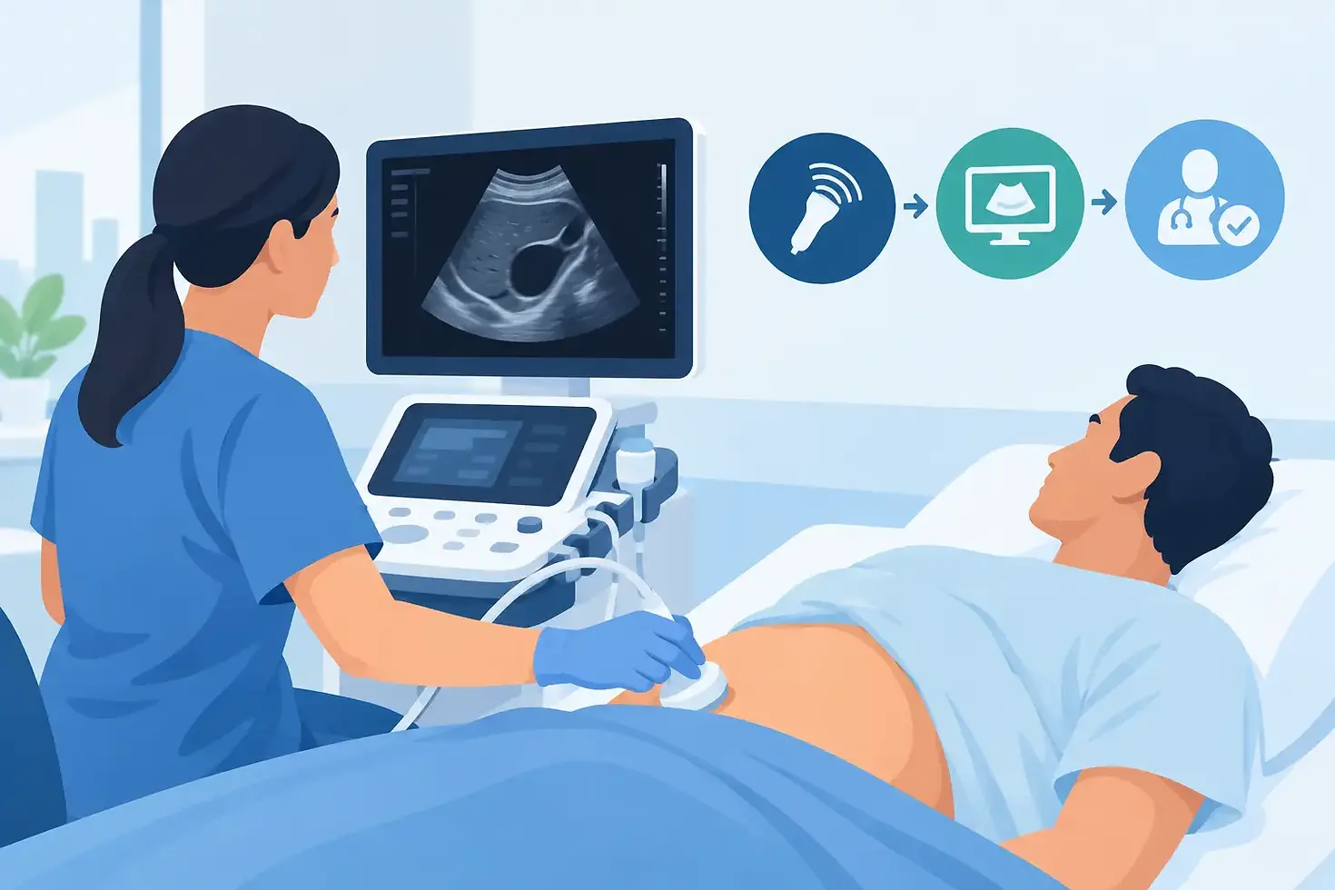
Bedside Ultrasound Workflow for Faster Decisions

A delayed ultrasound can hold up treatment, discharge planning, medication changes, and family communication. For patients who are homebound, medically fragile, in hospice, or living in a skilled nursing setting, transportation to an imaging center can create an additional clinical and logistical burden. A dependable bedside ultrasound workflow brings the exam to the patient while giving the care team a clear path from order to final interpretation.
The objective is not simply to place an ultrasound machine at the bedside. The process must protect patient identification, capture the right clinical question, obtain diagnostic images, route those images securely, and deliver a report that the ordering provider can act on. When each handoff is defined, mobile ultrasound becomes a practical extension of the care environment rather than another task for facility staff to manage.
Start the Bedside Ultrasound Workflow Before Arrival
A reliable exam begins with a complete, legible order. The ordering provider should identify the requested study, the patient’s full name and date of birth, relevant diagnosis or symptoms, provider information, and the best contact method for urgent findings. Clinical history matters. A request for a venous duplex, for example, is more useful when the sonographer knows whether the concern is new unilateral swelling, pain, a prior clot, or anticoagulant use.
Facility staff can prevent avoidable delays by confirming patient availability before the mobile team arrives. This includes verifying the room or home address, checking for isolation precautions, identifying whether a family member or representative needs to be present, and noting mobility limitations, contractures, wounds, or equipment that may affect positioning.
Match exam urgency to the clinical need
Not every bedside ultrasound requires the same response window. A routine surveillance study can be scheduled around the patient’s care plan. New pain, swelling, suspected urinary retention, or symptoms that may affect immediate treatment may require faster coordination. The ordering provider and mobile imaging partner should agree on what qualifies as urgent and how urgent findings will be communicated.
Urgency should not replace clinical judgment. If a patient has unstable vital signs, acute respiratory distress, signs of stroke, severe chest pain, or another potential emergency, emergency evaluation may be more appropriate than waiting for a mobile diagnostic exam. A bedside study supports care decisions, but it does not replace emergency response when a patient needs immediate intervention.
Prepare the Patient and the Care Space
At the bedside, patient comfort and image quality are closely connected. The sonographer needs enough access to position the patient safely, adjust lighting when necessary, and reach the body area being examined without pulling on oxygen tubing, feeding tubes, catheters, or other lines.
Staff should explain the exam in plain language before it begins. Patients and caregivers often worry that ultrasound will be painful or disruptive. In most cases, the exam uses a handheld transducer and gel on the skin. Some pressure may be needed to assess a vessel or obtain a better view, particularly during vascular studies, but the sonographer should adapt positioning and pressure to the patient’s tolerance whenever possible.
Patient identity must be confirmed using accepted identifiers before images are acquired. The order, patient identifiers, body part, laterality when applicable, and clinical indication should align. This step may feel routine, but it protects against one of the most damaging workflow failures: images or reports being attached to the wrong patient or wrong study.
Privacy is equally operational. In a shared room, staff may need to close a curtain, limit unnecessary traffic, and protect the patient’s dignity during abdominal, pelvic, or vascular imaging. In a private home, the same standards apply. A mobile setting should never lower expectations for confidentiality or respectful care.
Acquire Diagnostic Images at the Point of Care
The quality of a bedside ultrasound depends on the clinical question, the patient’s condition, and the ability to obtain required views. Limited mobility, obesity, edema, bowel gas, pain, dressings, and inability to follow breathing instructions can affect the study. These challenges do not mean the exam should not be attempted. They mean the care team should recognize when a mobile study can answer the question and when additional imaging may be needed.
A trained sonographer follows the protocol for the ordered study and documents the required measurements and images. For a general ultrasound, this may involve abdominal organs, kidneys and bladder, soft tissue, thyroid, or other requested anatomy. For vascular imaging, the protocol may include compressibility, Doppler waveforms, velocity measurements, and comparison of vessels as indicated by the study.
The sonographer’s role is image acquisition, not independent diagnosis. Clinical staff should avoid pressing the sonographer for preliminary conclusions at the bedside. Images need to be reviewed by the appropriate interpreting physician or credentialed specialist under the established reporting process. Keeping that boundary clear protects both the patient and the care team from acting on an incomplete impression.
Secure Image Transfer Is Part of Clinical Care
An exam is not complete when the probe leaves the patient’s skin. Images and technical information must be transferred through a secure, HIPAA-compliant process to the interpreting provider. Digital workflows reduce the risk of misplaced paperwork and allow authorized clinicians to access images and reports without waiting for physical delivery.
For facility teams, the practical question is simple: who receives the report, and how quickly can they see it? The answer should be established at intake, not after the exam. Specify the ordering provider, the facility contact, the preferred delivery method, and an alternate contact for after-hours or urgent communication.
D-RAD Mobile Imaging supports provider-friendly digital image access so care teams can incorporate diagnostic information into the patient record and next-step planning without unnecessary transport or administrative chasing.
Build a clear pathway for significant findings
A final report should document the findings, impression, interpreting provider, and any recommended follow-up within the provider’s scope. If a finding requires prompt attention, the communication pathway must be documented and followed. This may include direct notification to the ordering provider, nurse, facility supervisor, or designated on-call clinician.
Facilities should have a written process for receiving these communications. The person notified needs authority to escalate concerns, reach the provider, and document the action taken. A report sitting in a fax queue or generic inbox does not help a patient whose treatment plan needs to change.
Close the Loop After the Report Arrives
The final step in a bedside ultrasound workflow is clinical follow-through. The ordering provider reviews the report in context with the patient’s symptoms, examination, history, and other available tests. A normal or limited study may still require further evaluation if symptoms persist. An abnormal finding may lead to medication changes, specialist referral, emergency transfer, repeat imaging, or a revised plan of care.
For nursing facilities, home health agencies, and hospice organizations, report review should also trigger documentation updates. Care plans, medication administration records, incident notes, and family communications may need to reflect the provider’s direction. When this is done promptly, the ultrasound serves its purpose: it moves care forward.
This follow-up step is especially valuable for patients who would otherwise face a difficult trip to an imaging center. Avoiding transport can reduce discomfort, fall risk, caregiver burden, and disruption of established care routines. Still, bedside service is not the right answer for every imaging need. Some patients require advanced imaging, hospital resources, contrast studies, or immediate specialist evaluation. The right workflow recognizes those limits early.
Make Mobile Ultrasound Easier to Order Every Time
Consistency comes from standardizing the few details that commonly cause delays. Keep current order forms available to providers and facility staff. Maintain a list of preferred contacts and after-hours escalation instructions. Train staff to collect relevant history and prepare patients before the mobile team arrives. Review missed appointments, incomplete orders, and delayed reports to identify recurring gaps.
For organizations serving high-acuity or high-volume populations, a single mobile diagnostic partner can simplify coordination across ultrasound, X-ray, EKG, laboratory work, and other ordered services. The value is operational as much as clinical: fewer calls, fewer transportation arrangements, and fewer handoffs between separate vendors.
A well-run bedside ultrasound workflow gives providers the information they need while allowing vulnerable patients to remain where they are receiving care. The best next step is to define your order, communication, and escalation process before the next urgent study is needed.



Comments